
Phosphorylated phosphatidylinositol lipids, known as phosphoinositides, play a crucial role in controlling various cell activities like signaling, structural changes in the cell cytoskeleton, membrane dynamics and fluidity, and other processes. Phosphoinositides are crucial parts of cell membranes. The amounts of phosphoinositides are managed by specific enzymes called phosphoinositide kinases and phosphatases. Different types of phosphoinositides act as specific signals to interact with various phosphoinositide-binding proteins, influencing a wide range of cell activities such as protein transport, cell structure, the organization of the cell’s skeleton, ion channel operation, and gene expression.
 with hydroxyl groups at positions 3, 4, and 5 of the myoinositol head group, which serve as targets for phosphorylation. Right: Phosphoinositides with phosphorylation sites marked in red, and phosphate groups denoted by a circled P.")
Despite a growing body of evidence highlighting the crucial role of phosphoinositide signaling in various cellular functions, our understanding of the phosphoinositide composition in the retina remains limited.
In this study, Finkelstein and colleagues developed a non-radiolabeling methodology to profile and quantify phosphoinositides and phosphatidylinositol in the mouse retina. Additionally, they identified subcellular localization patterns of these phosphoinositides within photoreceptors in the mouse model.

The PI(4)P and PI(4,5)P2 lipid species were extracted from the mouse retina, and the absolute amounts of the measured lipids were approximately the same in dark-adapted and light-adapted mice.

The authors also analyzed changes in the ratio of PI(4)P/PI(4,5)P2 extracted from the retinas of dark- and light-adapted mice. No significant changes were detected in the ratios of these lipids in dark-adapted and light-adapted mice.
P and PI(4,5)P2 in retina. A The amounts of PI(4)P and PI(4,5)P2 in the retina is nearly the same. B The PI(4)P to PI(4,5)P2 ratio in the retinas of group Dark and group Light are nearly the same.")
PI(4)P and PI(4,5)P2 in retina. A The amounts of PI(4)P and PI(4,5)P2 in the retina is nearly the same. B The PI(4)P to PI(4,5)P2 ratio in the retinas of group Dark and group Light are nearly the same.
Finkelstein, Gospe et al., Cells 2020, 9, 1417; 10.3390/cells9061417
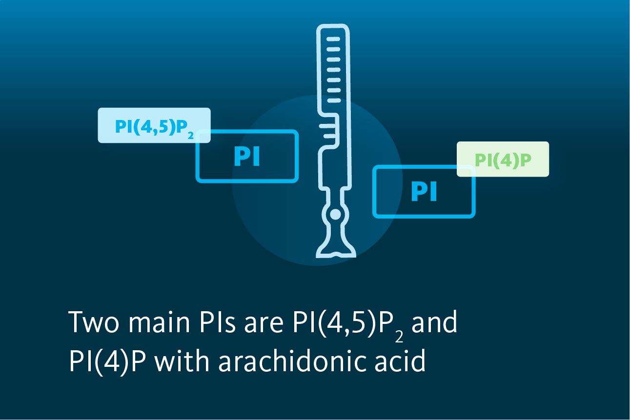
The phosphatidylinositol profile exhibited dominance by two species containing arachidonic acid: PI 18:0/20:4 (PI 38:4) and PI 16:0/20:4 (PI 36:4). These two species collectively constituted approximately 60% and 22% of the total retinal PI, respectively. The authors also identified trace amounts of PI species containing fatty acids with odd carbon numbers. Given the established understanding that mammals do not synthesize fatty acids with an odd number of carbons de novo, it is plausible that these fatty acids are sourced from a plant-based food diet.
The authors identified the prevailing in retina phosphorylated phosphatidylinositols as the 38:4 and 36:4 acylated species. Further, they observed comparable levels of PI(4)P and PI(4,5)P2. They estimated that phosphorylated inositides constitute approximately 7% of the total phosphatidylinositols containing 38:4/36:4 acyls. Lastly, the measured ratio of 38:4 to 36:4 acyl species remains largely unaffected by the status of inositol phosphorylation.
P, PI(4,5)P2), in dark-adapted mouse retinas.")
Retinal phosphoinositides. A Phosphatidylinositol profiles in mice retina with the schematic visualization of most abundant arachidonic acid-containing species. B Relative abundance of 36:4 and 38:4 PIs, along with their phosphorylated forms (PI(4)P and PI(4,5)P2) in dark-adapted mouse retinas.
Finkelstein, Gospe et al., Cells 2020, 9, 1417; 10.3390/cells9061417
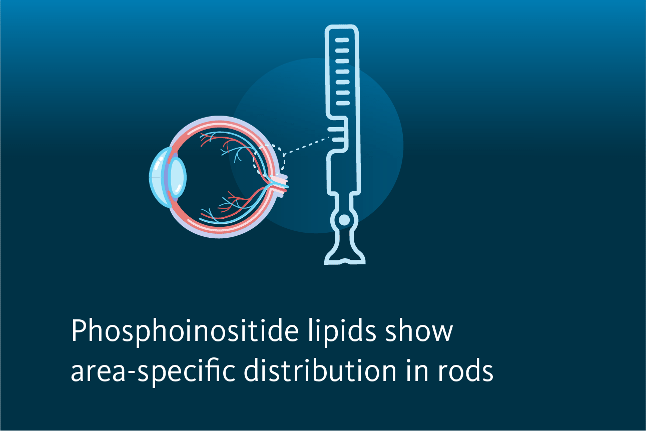
The scientists also explored the localization of phosphoinositides in rod photoreceptors by introducing GFP sensors for PI(4,5)P2 and PI(4)P into mouse rods via in vivo electroporation. They aimed to examine phosphoinositide’s presence in rod cell compartments.
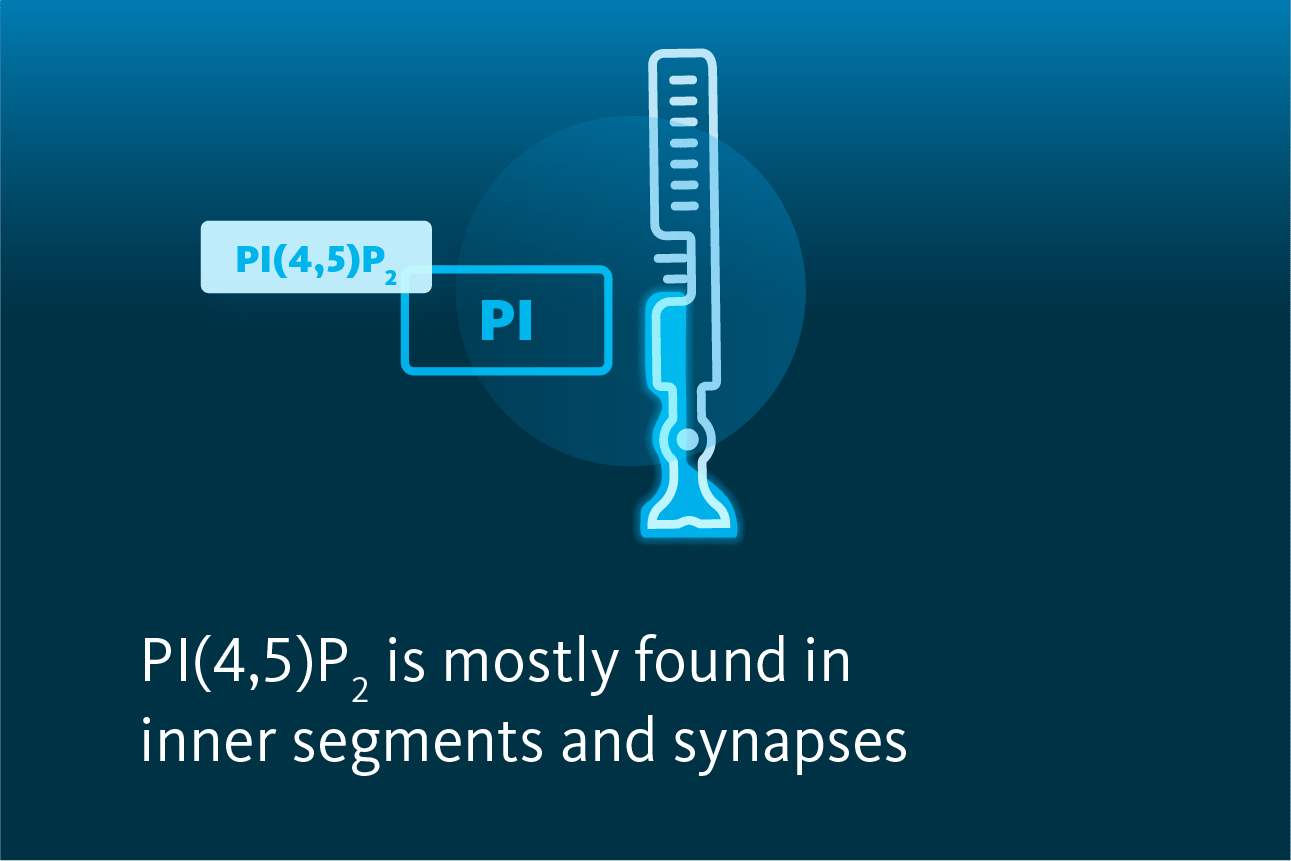
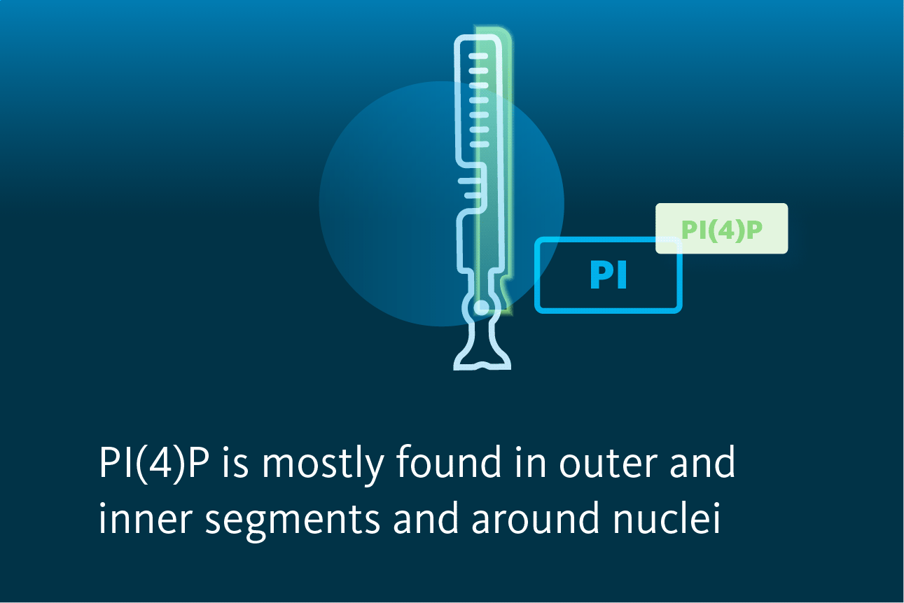
PI(4,5)P2 was found to be predominantly localized outside of outer segments, inner segments, and synapses. In contrast, the PI(4)P was present inside of outer segments, with a significant amount in inner segments and around nuclei. There was no distinct change in PI(4)P and PI(4,5)P2 localization between dark- and light-adapted mice.
P2 and PI(4)P in different rod compartments.")
To summarize, the authors analyzed PI(4)P and PI(4,5)P2 in the retinas of dark- and light-adapted mice and did not reveal any major differences. Lipidomics analysis showed that over 82% of the total phosphatidylinositide pool in the mouse retina comprises arachidonic acid-containing species, PI 38:4, and PI 36:4. This acyl composition was consistent for mono- and bisphosphate phosphorylated forms (PIP 36:4/38:4 and PIP2 36:4/38:4), constituting ~7% of total retinal phosphatidylinositol.
Using genetic phosphoinositide sensors, scientists studied the subcellular localization of PI(4,5)P2 and PI(4)P within rod photoreceptors. The main distinction in their distribution is that PI(4)P was present in the light-sensitive outer segment, while PI(4,5)P2 was mostly not present there yet present in photoreceptor synapses.
Lipotype Lipidomics technology can be used to characterize hundreds of phospholipids in various organs, tissues, cells, and organelles.
Not sure how this works for your case?
Let’s talk it through!

Lipotype products are provided for Research Use Only. They are not intended for clinical diagnostic purposes and must not be used to inform medical treatment decisions. The content of this article is for scientific and educational purposes only and should not be considered medical advice.