Home   Lipotype Lipidomics   Sphingolipids   Gangliosides   GD3 

GD3 Ganglioside

We provide MS-based lipid analysis of GD3 gangliosides in a variety of sample types and deliver results in as little as two weeks.

Details

Sphingolipids
Lipid Category
Gangliosides
Lipid Group
GD3 Ganglioside
Lipid Class

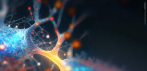
About the structure and biological function of GD3

Structure. 
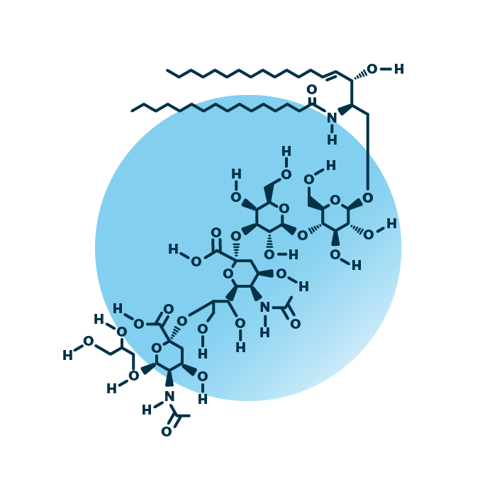
GD3 ganglioside lipids belong to the group of gangliosides within the sphingolipids. Their structure consists of a ceramide backbone linked to an oligosaccharide unit made of four sugar molecules. Two of them are sialic acid. The ceramide backbone contains two hydrocarbon chains: a long-chain base which is linked to a fatty acid via an amide bond. The fatty acid and the long-chain base can be of variable length, hydroxylated, and contain double bonds.

Function. 
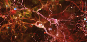
GD3 gangliosides are fundamental in neurogenesis. They bind to the EGF receptor resulting in the activation of a signaling cascade promoting cell proliferation. This is essential for stem cell self-renewal in the brain. GD3 lipids are critical for apoptosis and autophagy, programmed cell death and cellular “self-eating”. They also serve as precursors of more complex gangliosides. Further, GD3 levels are elevated in melanomas and neuroblastomas, and they are the main gangliosides in early human breast milk.

GD3 lipidomics analysis with
Lipotype

Structural detailsspecies level
Variants identified > 25
Approachuntargeted
Methodmass spectrometry
DeviceQ Exactive Orbitrap (280.000 Res)
Quantificationyes
Delivery time2-4 weeks
Lipidomics datapmol & mol%
Figuresincluded
1Glycosphingolipids includes:
DiHexCer, GM1, GM2, GM3, GM4, GD1, GD2, GD3, GT1, GT2, GT3, GQ1, Gb3, Gb4, Sulf

EXAMPLE STRUCTURE
GD3

An example for the molecular structure of ganglioside GD3, specifically GD3 34:1;2.

EXAMPLE
GD3 34:1;2

An example for the molecular structure of ganglioside GD3, specifically GD3 34:1;2.

ABOUT LIPOTYPE

Lipotype is the leading lipidomics service provider for industry and academia. Order your service, send your samples, get your data.

  • Thank you once again. The timing is extremely helpful to us.

    Susan Macdonald

    Aldeyra Therapeutics
  • Thank you very much. The results look fantastic!

    Stan Neufeld

    Friedrich Miescher Institute
  • The high-speed turn around when under revision really helped us!

    Rose E Goodchild

    KU Leuven
  • We highly appreciate the personal support and consultation.

    Rainer Voegeli

    DSM Skin Biology
  • We have been quite impressed. The results clearly indicate a difference.

    Pietro De Camilli

    Yale University
  • Cooperating with Lipotype, I enjoy custom packages and great support!

    Olga Ramich

    DIfE
  • After moving from one continent to another, I still prefer Lipotype!

    Julie Jacquemyn

    University of Alberta
  • Thank you very much for my new and shiny lipidomics data set!

    Ilya Levental

    University of Virginia
  • Lipotype Lipidomics is instrumental to our studies of membranes.

    Christopher J Stefan

    University College London
  • Lipotype is truly dedicated to supporting scientists in their research.

    Anonymous

    Anonymous

Contact Lipotype