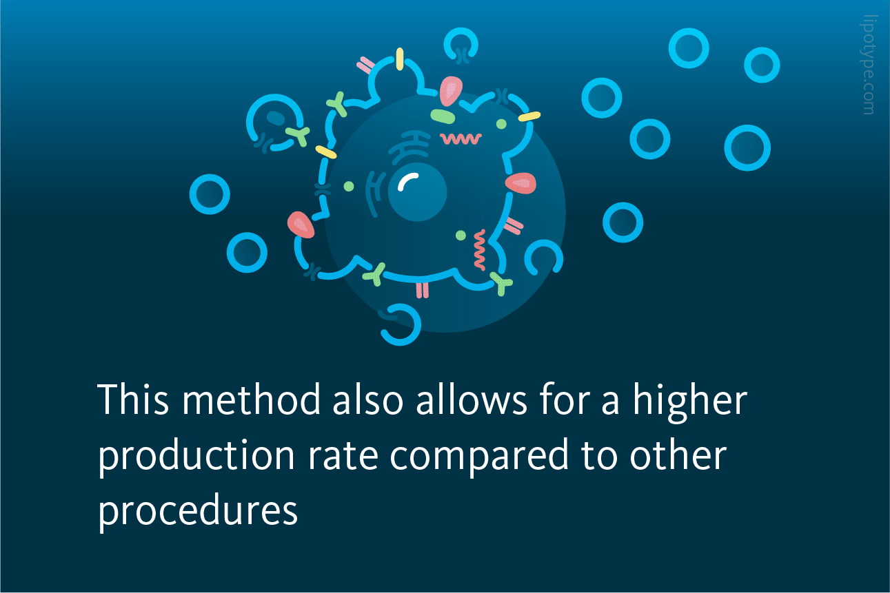
EXTRACELLULAR vesicles (EVs) are tiny membranous structures released by cells. EVs contain various cell-specific materials like lipids, proteins, nucleic acids, metabolites, and organelles. EVs are divided into two main types: exosomes and ectosomes. Exosomes are small (40-150 nm) and come from inside cells, released through a continuous process called exocytosis. Ectosomes, on the other hand, form directly from the cell’s plasma membrane and include different vesicles like microvesicles, apoptotic bodies (ApoBDs), apoptotic extracellular vesicles (ApoEVs), and large oncosomes. Ectosomes can vary in size from 50 nm to over 1000 nm.
Extracellular vesicles have unique characteristics inherited from their parent cells. They can travel to distant cells within living organisms, get taken up by them, and change how those cells behave. Extracellular vesicles are used for delivering drugs in experiments on both cells and animals. However, using EVs for treatments has been challenging because producing them in large quantities is rather slow and requires a lot of donor cells. Furthermore, isolating, purifying, and separating the produced EVs adds complexity to the process. Complicating matters further, different laboratories use varying protocols and experimental conditions. To speed up research and the use of EV-based therapies, EV societies are creating guidelines to standardize production procedures.

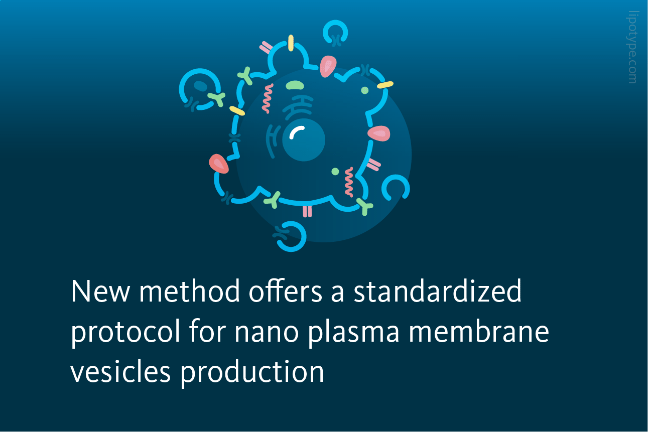
A study by Alter and colleagues aimed to overcome limitations in standard laboratory protocols for preparing EVs. They sought to develop an efficient method that could produce consistent EVs with high reproducibility. To do this, they used Giant Plasma Membrane Vesicles (GPMVs), which are created through chemical stressors that induce cell apoptosis and membrane blebbing. These GPMVs were then considered as a starting material for making EVs.
Next, they reduced the size of these GPMVs through a process called extrusion, resulting in nano Plasma Membrane Vesicles (nPMVs). The researchers conducted a comprehensive analysis of nPMVs, comparing them to native EVs obtained from the same donor cell line. They looked at various aspects of produced EVs, including production yields, physico-chemical properties, morphology, molecular composition including lipidomics, and other functions.

In terms of storage stability, samples stored at 4°C remained stable for up to 1 month after production. The colloidal stability of both nPMVs and native EVs was similar under these conditions. However, nPMV samples stored at room temperature showed an increase in hydrodynamic diameter and polydispersity index – parameters commonly used to characterize EVs – after 1 month of storage. The authors determined the production yields and rates for both the nPMV and native EV preparation methods using particle concentrations, sample volumes, and cell numbers at the initial stage of particle production. They found that the production rate for nPMVs was 1100–1386 particles per cell per hour, whereas, for the native EV preparation method, it was 5–6 particles per cell per hour (p ≤ 0.01).
Lipids play a crucial role in the entry of enveloped viruses and parasites into cells, and they are also believed to be important for exosomes in cell entry. Because of this, the authors decided to analyze the lipid composition of Huh7 cells and Huh7 nPMVs using mass spectrometry-based lipidomics. They discovered that nPMVs were statistically significantly enriched in certain lipids like phosphatidylserine (PS), sphingomyelin (SM), and phosphatidylcholine (PC) when compared to the donor cells (p ≤ 0.01). Specifically, PS content in nPMVs was increased comprising 14.3 mol% of the total lipid content compared to 3.7% in the donor cells. On the other hand, lipids like phosphatidylethanolamine, phosphatidylglycerol, and phosphatidylinositol were present in lower amounts in nPMVs compared to donor cells (p ≤ 0.05). Di- and triglycerides were found in host cells but were absent in nPMVs (p ≤ 0.001). The lack of diglyceride and triglyceride in nPMV indicates the absence or minimal presence of lipid droplets or similar lipoparticles.

Selected lipid classes profiles of Huh7 nPMVs and parent cells. Lipidomic analysis of Huh7 nPMVs and Huh7 parent cells. Values are means ± SD, n = 3. Levels of significance: *p ≤ 0.05, **p ≤ 0.01, ***p ≤ 0.001. Huh7 cells are liver cancer cell culture.
Alter et al., Commun biol (2023) 6, 478, 10.1038/s42003-023-04859-2
The lipidomic analysis revealed some interesting findings in the comparison between Huh7 nPMVs and their donor cells. nPMVs were found to be enriched in lipids like PC, PS, SM, and cholesterol esters compared to their donor cells. However, it’s worth mentioning that this observation contradicts most of the existing literature, which typically shows an enrichment of cholesterol, PS, SM, and glycosphingolipids in EVs from various cell lines. These differences can likely be attributed to the distinct origins and preparation methods of nPMVs and native EVs.
The authors noted a nearly four-fold higher PS content in nPMVs compared to donor cells, whereas most EV studies report only a one- to three-fold enrichment. The PS content in ApoBDs and exosomes can vary depending on the vesicle’s cellular origin or the donor cell’s phenotype. However, what’s more relevant for their biological properties is the location of PS in the nanoparticle’s membrane rather than the overall PS content. PS exposed on the outer surface of nanoparticles provides a strong “eat-me” signal for receptors. This process is exploited by membrane-enveloped viruses and parasites as a crucial factor for their entry into host cells. The authors demonstrated that PS was indeed exposed on the surface of both nPMVs and native EVs. This suggests that nPMVs, derived from GPMVs (ApoBDs), likely consist primarily of ApoEVs.

The EV preparation method described by the authors for nPMVs addresses several limitations of existing EV preparation methods, including efficiency, uniformity, and reproducibility. This method outperforms conventional EV preparation methods in terms of production rate and yield by at least one to two orders of magnitude, making it possible to produce EVs with consistent size and reproducibility more quickly. A systematic comparison between nPMVs and native EVs from the same cells revealed biologically relevant differences in proteomes and lipidomes, which can be attributed to the distinct origins and preparation methods of these vesicles. The EV preparation technology described in this study could serve as a foundation for developing therapeutic agents, either using nPMVs independently for immunotherapy or in combination with low-molecular-weight drugs or therapeutic biomolecules.

Lipotype Lipidomics technology can be used to characterize the membrane lipidomics changes triggered by various factors, such as exposure to different temperatures or various diets.
Do our methods apply to your samples?
Let’s figure that out!
Lipotype products are provided for Research Use Only. They are not intended for clinical diagnostic purposes and must not be used to inform medical treatment decisions. The content of this article is for scientific and educational purposes only and should not be considered medical advice.