
by Dominika Bezdeková
LIPIDS are essential for cell membranes and energy storage, but when they accumulate in the wrong places – a condition known as ectopic lipid deposition – they can disrupt cellular function. This disruption alters mitochondrial dynamics, contributing to the development of insulin resistance and obesity. Among these lipids, sphingolipids have emerged as essential players in metabolic regulation.
Sphingolipids are a class of lipids characterized by a sphingoid base backbone, a long-chain amino alcohol. Ceramides, the simplest sphingolipids, consist of a fatty acid attached to this backbone. These ceramides increase during obesity, promote insulin resistance, and act as key mediators in cellular signaling pathways and lipid metabolism in general. Moreover, they serve as precursors for more complex sphingolipids, such as sphingomyelins (containing a phosphorylcholine head group) or glycosphingolipids (containing one or more sugar molecules).

Dietary components influence ceramide production and buildup in the body. The Western diet, known for its high intake of energy-dense foods rich in long-chain saturated fatty acids (LCSFAs), especially palmitic acid, contributes to ceramide formation. These fatty acids trigger inflammation, activating key enzymes that drive ceramide production. LCSFAs may reduce mitochondrial oxidative capacity, leading to fatty acid accumulation, which further supports ceramide synthesis. Besides palmitic acid, fructose can also increase ceramide levels. The Mediterranean diet is rich in monounsaturated fats (MUFA) and omega-3 polyunsaturated fatty acids (PUFA). It can lower ceramide levels by enhancing mitochondrial oxidative function, improving metabolic flexibility, and reducing inflammation. Omega-3 fatty acids and inulin also support gut microbiota health by increasing its diversity and promoting the production of beneficial short-chain fatty acids.
, especially palmitic acid, contributes to ceramide formation. These fatty acids trigger inflammation, activating key enzymes that drive ceramide production. LCSFAs may reduce mitochondrial oxidative capacity, leading to fatty acid accumulation, which further supports ceramide synthesis. Besides palmitic acid, fructose can also increase ceramide levels. The Mediterranean diet is rich in monounsaturated fats (MUFA) and omega-3 polyunsaturated fatty acids (PUFA). It can lower ceramide levels by enhancing mitochondrial oxidative function, improving metabolic flexibility, and reducing inflammation. Omega-3 fatty acids and inulin also support gut microbiota health by increasing its diversity and promoting the production of beneficial short-chain fatty acids.")
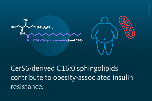
This study specifically focused on C16:0 sphingolipids and their ceramide precursors derived from two enzymes: Ceramide Synthase 5 (CerS5) and Ceramide Synthase 6 (CerS6). These sphingolipids include 16-carbon fatty acid ceramides produced by these enzymes, along with all their sphingolipid derivatives. Although both CerS5 and CerS6 generate the same type of ceramides, these lipids differ significantly in their functions and effects due to their distinct tissue distribution and cellular localization.
C16:0 sphingolipids derived from CerS5 and CerS6 enzymes are present in the liver and brown adipose tissue – key hubs of energy metabolism. However, CerS6-derived sphingolipids play a more prominent role in regulating energy metabolism, by inhibiting the breakdown of fatty acids into smaller molecules.

In this study, Philipp Hammerschmidt and colleagues compared the effects of deleting CerS6 and CerS5, enzymes producing C16:0 ceramide lipids, in obese mice fed a high-fat diet. Mice lacking the CerS5 gained weight to the same extent as controls (unmodified wild-type fed a high-fat diet), while those with CerS6-deficiency showed reduced weight gain. Moreover, the CerS6-lacking mice also exhibited lower adiposity, decreased diet-induced liver weight, and reduced hepatic lipid accumulation – effects not observed in CerS5-deficient mice.
Although hepatic C16:0 ceramide levels were significantly reduced in both experimental groups, only the CerS5-deficient mice showed additional reductions in C22:0 and C24:0 ceramides. These results highlight that while both enzymes influence hepatic ceramide levels, only CerS6 deficiency protects from the metabolic effects of high-fat diet feeding – diet-induced obesity and obesity-associated insulin resistance.

Deficiency of CerS6, but not CerS5, protects from diet-Induced obesity, hepatic lipid accumulation, insulin resistance, and mitochondrial dysfunction. Body weights of A CerS6 and B CerS5 deficient mice compared to control mice. Quantification of fat content relative to body weight in C CerS6 and D CerS5 deficient mice compared to control mice. Ceramide levels in the liver of E CerS6 and F CerS5 deficient mice compared to control mice. Data are represented as mean ± SEM.
Hammerschmidt et al. Cell, 177.6: 1536-1552 (2019), 10.1016/j.cell.2019.05.008
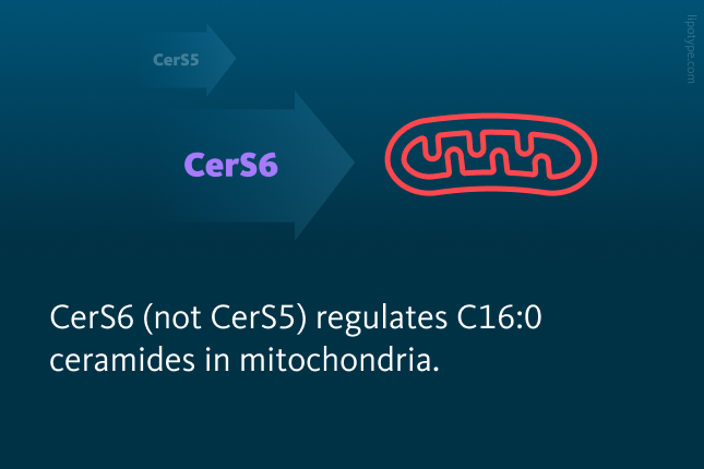
C16:0 ceramides and their sphingolipid derivatives are regulated differently by CerS enzymes across cellular compartments. In mitochondria and mitochondria-associated membranes (MAMs), their levels are specifically regulated by CerS6, but not CerS5. These cellular compartments are critical for energy production and lipid exchange, making them hotspots for ceramide-induced mitochondrial fragmentation.
By deleting the CerS6 enzyme, harmful C16:0 ceramide levels in mitochondria and MAMs can be significantly reduced, restoring mitochondrial structure and function. In contrast, CerS5 deficiency does not alter mitochondrial ceramide levels, even though the overall hepatic C16:0 ceramide content is also reduced.
 regulates mitochondria-associated membranes (MAM) and mitochondrial C16:0 ceramide pools morphology in the liver. Ceramide profile of liver homogenate (top row), mitochondria-associated membranes (middle row), and mitochondrial fractions (bottom row) of clean diet and high-fat diet-fed mice. Ceramide profile of liver subcellular fractions of high-fat diet-fed CerS5 (in blue) deficient and CerS6 (in red) deficient mice compared to control mice.")
CerS6 (not CerS5) regulates mitochondria-associated membranes (MAM) and mitochondrial C16:0 ceramide pools morphology in the liver. Ceramide profile of liver homogenate (top row), mitochondria-associated membranes (middle row), and mitochondrial fractions (bottom row) of clean diet and high-fat diet-fed mice. Ceramide profile of liver subcellular fractions of high-fat diet-fed CerS5 (in blue) deficient and CerS6 (in red) deficient mice compared to control mice.
Hammerschmidt et al. Cell, 177.6: 1536-1552 (2019), 10.1016/j.cell.2019.05.008
In obesity, mitochondria structure and function are often impaired due to a phenomenon known as mitochondrial fragmentation. As found out by this study, C16:0 sphingolipids derived from CerS6 directly contribute to this fragmentation process by interacting with a sphingolipid-binding protein called mitochondrial fission factor (Mff). The interaction then promotes mitochondrial dysfunction, aggravating insulin resistance and diet-induced obesity.
This study showed that by invalidating the interaction and reducing C16:0 ceramide synthesis through CerS6 deficiency, mitochondrial fragmentation could be mitigated, improving mitochondrial dynamics. The experiments demonstrated in both in vitro cellular and in vivo models, showed that insulin sensitivity, glucose, and pyruvate tolerance were markedly improved, offering protection against obesity and diabetes.

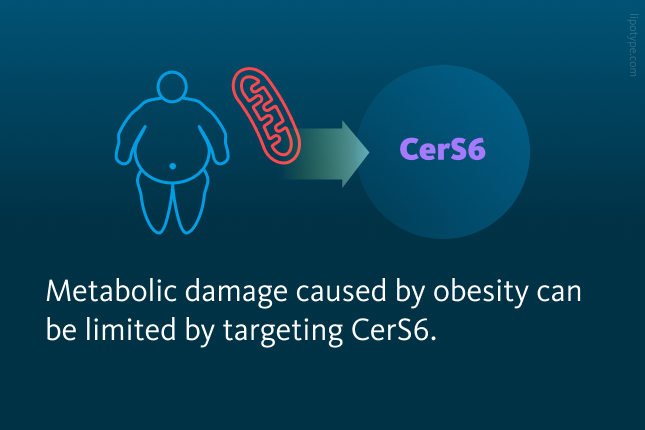
This study revealed a distinct specificity of C16:0 sphingolipids derived from CerS6 and CerS5 in diet-induced obesity and insulin resistance. CerS6, unlike CerS5, regulates C16:0 ceramide levels in mitochondria and MAMs, directly impacting mitochondrial dynamics. By targeting the CerS6 enzyme and reducing its derived sphingolipids, researchers demonstrated the potential to alleviate mitochondrial dysfunction, reduce obesity-induced metabolic stress, and improve insulin resistance. These findings highlight the therapeutic potential of CerS6 inhibition in combating obesity and diabetes.

Lipotype Lipidomics technology supports researchers in performing a comprehensive analysis of lipid profiles, helping to identify specific changes. By gaining precise lipidomic insights, researchers contribute to developing prevention options and therapies for obesity, insulin resistance, and related conditions.
Do you have any questions?
We can answer them!

Lipotype products are provided for Research Use Only. They are not intended for clinical diagnostic purposes and must not be used to inform medical treatment decisions. The content of this article is for scientific and educational purposes only and should not be considered medical advice.