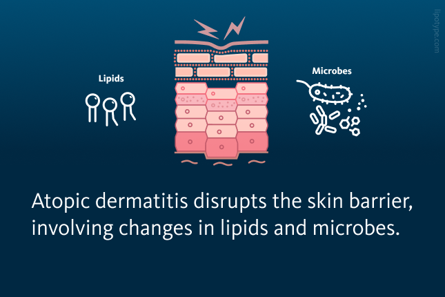
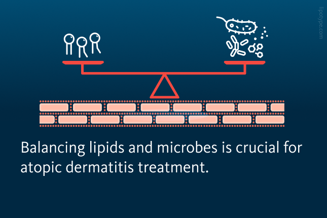
THE skin’s protective barrier prevents water loss and defends the body against external threats like allergens and microbes. This barrier depends on mature keratinocytes, a lipid-rich extracellular matrix, and a balanced community of resident microbes – our normal, healthy microbiota. When this balance is disturbed, along with other factors like genetic predisposition and immune responses, it can contribute to skin disorders such as atopic dermatitis, the most common form of eczema. While microbial and lipid imbalances are not the sole cause of atopic dermatitis, they play a key role in disease development and progression.
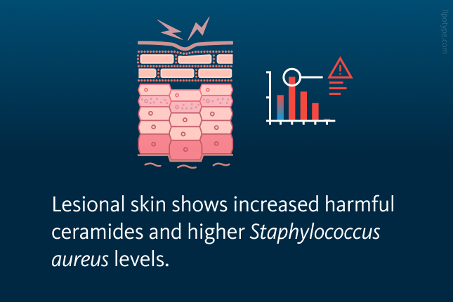
In healthy skin, diverse microbial communities coexist peacefully with the skin’s lipids to support barrier function. In contrast, atopic dermatitis is characterized by an overgrowth of potentially harmful bacteria like Staphylococcus aureus (S. aureus) and reduced microbial diversity, alongside changes in lipids such as short-chain fatty acids, specific ceramides, and sphingomyelins that normally maintain barrier integrity. Research linking lipid and microbiome shifts shows that certain long-chain fatty acids in atopic dermatitis may correlate with bacterial groups like Corynebacterium, highlighting the need to study the skin microbiota and lipids together to better understand skin health and disease.

In this study, Bhattacharyya and colleagues examined how skin surface lipids and microbial communities interact in atopic dermatitis. To do this, researchers combined shotgun lipidomics, which provides broad coverage of lipid species, with microbial DNA sequencing. Tape-strip samples were collected from lesional and non-lesional skin of atopic dermatitis patients as well as from matched sites in healthy individuals, allowing them to directly compare lipid and microbiome profiles across groups.
This study included sixteen adults with atopic dermatitis and sixteen age- and sex-matched healthy controls. Atopic dermatitis patients avoided topical treatments before sampling, and tape strips were taken primarily from the hands or legs, with some from the torso. From each patient, lesional and non-lesional skin sites were sampled in parallel, and healthy controls were sampled at equivalent anatomical sites. Lipids were extracted for comprehensive profiling, and microbial DNA was sequenced to characterize community composition. In addition, selected tape strips were used in culture-based assays to explore how specific skin bacteria interact with components collected from the skin surface.

To explore whether epidermal lipids and skin commensal bacteria (microbes that normally live on the skin without causing harm) are linked, researchers analyzed lipid and microbiome profiles from tape-strip samples of atopic dermatitis patients and matched healthy controls. Lesional atopic dermatitis skin showed a strong dominance of cholesterol and had higher overall lipid levels than non-lesional and healthy skin. Cluster analysis revealed that several ceramide subclasses, particularly non-hydroxy-sphingosines (NS), non-hydroxy-dihydrosphingosine (NdS), non-hydroxy-6-hydroxyshingosines (NH), and alpha-hydroxy-sphingosine (AS) were most abundant in atopic dermatitis samples, with levels decreasing from lesional to non-lesional to healthy skin. In contrast, omega-hydroxy-6-hydroxysphingosine (EOH), omega-hydroxy-sphingosine (EOS), alpha-hydroxy-dihydrosphingosine (AdS), alpha-hydroxy-6-hydroxysphingosine (AH), and alpha-hydroxy-phytosphingosine (AP) ceramides increased along this gradient, reaching their lowest levels in lesional sites.
Across all samples, lipid diversity was lowest in lesional atopic dermatitis skin and gradually increased in non-lesional and healthy skin. Variability in lipid profiles was influenced not only by disease status but also by age, sex, and sampling location.
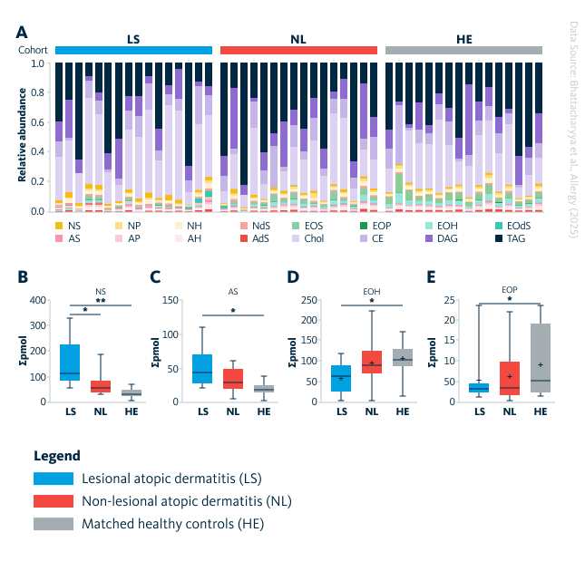
Overview of lipid profiles in lesional atopic dermatitis (LS), non-lesional skin (NL), and site-matched healthy controls (HE). A Overall lipid class distribution is shown as percentage for each group. B-E Box plots display four lipid classes that differed significantly across groups (NS, AS, EOH, and EOP). *, p-value < 0.05; **, p-value < 0.01. AdS, alpha-hydroxy-dehydrosphingosine; AS, α-hydroxy-sphingosine; AH, α-hydroxy-6-hydroxysphingosine; AP, α-hydroxy-phytosphingosine; CE, cholesteryl esters; Chol, cholesterol; DAG, diacylglycerols; EOdS, ω-hydroxy-dehydrosphingosine; EOH, ω-hydroxy-6-hydroxysphingosine; EOP, ω-hydroxy-phytosphingosine; EOS, ω-hydroxy-sphingosine; NdS, non-hydroxy-dehydrosphingosine; NH, non-hydroxy-6-hydroxysphingosine; NP, non-hydroxy-phytosphingosine; NS, non-hydroxy-sphingosine; TAG, triacylglycerols.
Bhattacharyya et al., Allergy, 2025, 0:1-14, 10.1111/all.70028
Microbiome analysis of the same skin sites demonstrated increased microbial abundance in both lesional and non-lesional atopic dermatitis skin compared to healthy controls, particularly of Staphylococcus species. Unlike the lipid profile, microbial diversity was highest in lesional skin. Age and sex had minimal effects, whereas sampling site and lesion type accounted for the majority of variance. Diversity analysis indicated significant differences across all groups. Twelve key microbial species were identified, including an enrichment of S. aureus in lesions and a reduction of S. melonis, Arthrobacter russicus, and S. aquatilis. Interestingly, S. hominis and Finegoldia magna were more abundant in non-lesional skin compared to healthy controls.
, non-lesional skin (NL), and matched healthy controls (HE). Relative abundance of the ten most prevalent bacterial species is shown for each group.")
Microbial composition in lesional atopic dermatitis (LS), non-lesional skin (NL), and matched healthy controls (HE). Relative abundance of the ten most prevalent bacterial species is shown for each group.
Bhattacharyya et al., Allergy, 2025, 0:1-14, 10.1111/all.70028
To better understand the role of Staphylococcus species beyond S. aureus in atopic dermatitis, species were categorized based on known pro-inflammatory or protective effects. Inflammatory species (S. aureus, S. caprae, S. petrasii) were compared to commensals with potential protective roles or those commonly found on healthy skin without evidence of pro-inflammatory activity (S. warneri, S. equorum, S. capitis, S. hominis, S. epidermidis, S. saccharolyticus). Their relative abundances were analyzed across lesional, non-lesional, and healthy skin. The ratio of inflammatory to protective species was significantly higher in lesional skin compared to non-lesional and healthy skin, with an even stronger difference observed when specifically comparing the S. aureus to S. hominis ratio.

Correlation analyses linked lipid groups with microbial families and species, and network centrality measures were used to assess how connected these lipid-microbe relationships were. Because Staphylococcus species drove most group differences, networks were built around them. Non-lesional and healthy skin showed dense, interconnected networks, whereas lesional skin displayed sparse, isolated modules. Closeness increased, and radiality decreased from lesional to healthy skin, indicating more integrated networks in healthier tissue. In healthy skin, S. aureus showed many correlations with ceramides and glycerolipids, while in non-lesional skin, these correlations were fewer and often negative. S. hominis showed limited interactions in lesional skin but increasing diversity toward healthy skin. Overall, lesional skin formed more microbe-centric and weakly connected lipid interaction networks.
To better understand microbial-lipid interactions under controlled conditions, lipid profiles from tape-stripped reconstructed human epidermis (RHE) models mimicking atopic dermatitis were compared to those from lesional patient samples. Among 249 lipids identified in RHE, 180 overlapped with 392 lipids from lesional skin samples. Both RHE and lesional skin shared similar distributions of ceramide lipids and glycerol esters, supporting the relevance of RHE as an in vitro model for studying lipid-microbe interactions in atopic dermatitis.
 skin and reconstructed human epidermis (RHE). A Overlap between lipids detected in RHE samples and those identified in lesional AD skin. B Distribution of selected lipid classes and ceramide subclasses in RHE compared with lesional AD samples.")
Lipid profiles in atopic dermatitis (LS) skin and reconstructed human epidermis (RHE). A Overlap between lipids detected in RHE samples and those identified in lesional AD skin. B Distribution of selected lipid classes and ceramide subclasses in RHE compared with lesional AD samples.
Bhattacharyya et al., Allergy, 2025, 0:1-14, 10.1111/all.70028
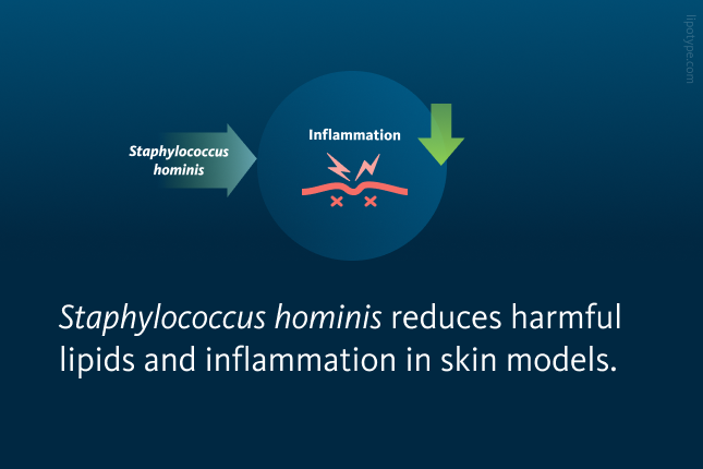
RHE models mimicking atopic dermatitis shared key lipid features with lesional skin, allowing a reduced microbe-lipid network to be built. Within this network, S. hominis emerged as a candidate of interest because it negatively correlated with the lesional-enriched ceramide NdS 18:0;2/24:0;0 and positively with certain diacylglycerols. In atopic dermatitis-like RHE, adding S. hominis improved skin structure, reduced this harmful ceramide, and broadly shifted lipid composition. Direct contact assays confirmed that S. hominis can lower NdS 18:0;2/24:0;0. In vivo, S. hominis associated with better skin barrier function (lower transepidermal water loss), whereas this ceramide associated with worse barrier integrity. RNA-seq showed that S. hominis partially rescued atopic dermatitis-related inflammatory and lipid-metabolic gene changes, supporting a dual role in reducing inflammation and promoting skin barrier homeostasis.
Overall, the data show that Staphylococcus hominis plays a protective and restorative role in atopic dermatitis by counteracting harmful lipids, improving skin architecture, and affecting inflammation. Together with prior evidence of its antimicrobial and immunomodulatory activity, these findings position S. hominis as a promising candidate for microbiome-based therapies and highlight the broader importance of microbe-lipid interactions in maintaining skin barrier health.
Lipotype Skin Lipidomics technology offers support to cosmetics researchers and dermatologists by facilitating the product development, for example, moisturizers and measuring differences in effects of these products on different types of skin. Detailed lipidomics analysis helps to develop innovative moisturizers and other skin care products.
Do you have any questions?
We can answer them!

Lipotype products are provided for Research Use Only. They are not intended for clinical diagnostic purposes and must not be used to inform medical treatment decisions. The content of this article is for scientific and educational purposes only and should not be considered medical advice.