
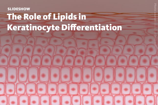
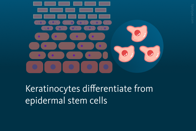
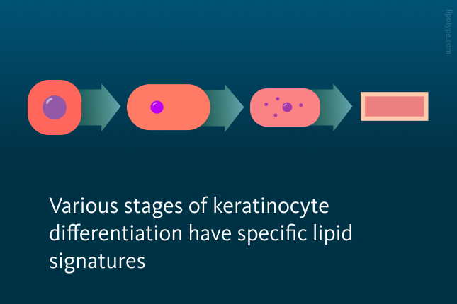
The epidermis, the outer layer of skin, is made up mainly of keratinocytes. Keratinocytes are a type of cell that migrate from the basal layer (stratum basale), where skin stem cells are found, and gradually move upward through the spinous (stratum espinosum) and granular layers (stratum granulosum) to the surface (stratum corneum). As they migrate, keratinocytes go through the final stages of differentiation, secreting lipid-rich lamellar bodies. Eventually, these cells lose their nuclei, transforming into corneocytes, which are bound by lipid layers in a structure resembling bricks and mortar.

Matteo Rudan and colleagues from King’s College London aimed to explore how lipids influence keratinocyte differentiation. They conducted a lipidomic analysis of the cultured human epidermal keratinocytes with disrupted lipid metabolism, examining the link of lipid metabolism to the start of differentiation. They identified lipid species that prompted cells to exit the epidermal stem cell compartment.

Differentiation of primary human keratinocytes, triggered by single-cell suspension, can be inhibited with a protein kinase C inhibitor (PKCi). To investigate lipid changes in suspension culture, the authors applied shotgun lipidomics to compare lipid profiles between control cells and those treated with PKCi.
Unsupervised clustering based on lipid abundance revealed the main sample groups: adherent cells, differentiated cells, committing cells, and samples with inhibited differentiation. Lipid analysis revealed a significant buildup of ceramides and hexosylceramides as keratinocytes differentiated in suspension. Such an increase in these lipids is also seen during epidermal differentiation in vivo. This accumulation was inhibited with PKCi.
. Error bars representing standard deviations (SDs).")
Ceramides and hexosylceramides changes in keratinocytes. Changes in ceramides and hexosylceramides at the class level during keratinocyte differentiation induced by suspension (in pmol% of total). Error bars representing standard deviations (SDs).
Vietri Rudan et al. PNAS, 117(36), 22173-22182, (2020), 10.1073/pnas.2011310117
Principal component analysis (PCA) separated the adherent cells from all other samples along the first component, while the differentiated cells were distinct along the second. However, committing and inhibited cells were not clearly separated. To pinpoint key lipid species that accumulated during commitment and differentiation, samples were classified into four categories—“adherent,” “committing,” “differentiated,” and “inhibited”—and analyzed using sparse partial least squares regression (sPLS) with discriminant analysis (DA) to identify the main lipids driving separation between groups.
 of sample variation across time points. Color used in infographics: green – Adherent (00 h), blue – Committing (04 and 08 h), orange – Differentiated (12 and 24h), grey – Inhibited (all time points).")
Principal Component Analysis (PCA) of sample variation across time points. Color used in infographics: green – Adherent (00 h), blue – Committing (04 and 08 h), orange – Differentiated (12 and 24h), grey – Inhibited (all time points).
Vietri Rudan et al. PNAS, 117(36), 22173-22182, (2020), 10.1073/pnas.2011310117
In the sPLS regression, the first component separated adherent samples, the second isolated differentiated samples, and the third distinguished committing samples. Lipid species contributing to components 2 and 3 helped identify specific molecules enriched in committing and differentiated samples, with a total of 145 lipids accumulating. These findings indicate that during suspension-induced differentiation, keratinocytes undergo substantial changes in lipid composition, accumulating specific lipid species.
, blue – Committing (04 and 08 h), orange – Differentiated (12 and 24h), grey – Inhibited (all time points).")
Segregation of samples across three components using sPLS. Color used in infographics: green – Adherent (00 h), blue – Committing (04 and 08 h), orange – Differentiated (12 and 24h), grey – Inhibited (all time points).
Vietri Rudan et al. PNAS, 117(36), 22173-22182, (2020), 10.1073/pnas.2011310117
The authors demonstrated that disrupting lipid metabolism in primary human keratinocytes can affect terminal differentiation. Lipidomics analysis pointed to potential lipid regulators of this process. To precisely identify which lipid species influence differentiation, researchers conducted a detailed lipidomic analysis of cells transfected with siRNA targeting ELOVL1 or SLC27A1 (involved in fatty acid elongation and lipid transport) at 24-, 48-, and 72-hours post-transfection. To identify lipidome changes in knockdown cells, they performed a separate sPLS-DA analysis on the 48- and 72-hour samples.
195 unique lipid species enriched in siELOVL1 cells were identified at either time point and 148 species in siSLC27A cells. The authors then compared lipids enriched in committing and differentiated cells with those in ELOVL1- or SLC27A1-knockdown keratinocytes. An overlap of 26 and 16 potential differentiation inducer lipids for ELOVL1 and SLC27A1 knockdown was detected. These lipids included sphingolipids (ceramides and hexosylceramides) and glycerophospholipids (phosphatidylcholines and phosphatidylserines).
. Statistical significance is determined by one-way ANOVA with Dunnett’s multiple comparisons test (*P < 0.05, **P < 0.01, ***P < 0.001, ****P < 0.0001) relative to the control.")
Lipid molecules promote keratinocyte differentiation in culture. Overlap of discriminant lipid sets enriched during ELOVL1 knockdown, SLC27A1 knockdown, and suspension-induced differentiation of keratinocytes. Keratinocyte response to ceramides and glucosylceramides is visualized in plots. Error bars represent standard deviations (SDs). Statistical significance is determined by one-way ANOVA with Dunnett’s multiple comparisons test (*P < 0.05, **P < 0.01, ***P < 0.001, ****P < 0.0001) relative to the control.
Vietri Rudan et al. PNAS, 117(36), 22173-22182, (2020), 10.1073/pnas.2011310117
The authors checked if two selected ceramides and two selected glucosylceramides from the intersection sets could induce keratinocyte differentiation. All candidate lipids increased the number of irreversibly differentiated keratinocytes.
Overall, this study demonstrates that the lipid profile of human keratinocytes is crucial for their differentiation, with effects traceable to specific lipid subspecies. Lipidomics analysis identified lipid species that, when added to cultured keratinocytes, actively induce differentiation. These findings highlight the role of individual lipid species in regulating cellular functions, setting the stage for further investigation into lipids’ role in epidermal differentiation.
Lipotype Lipidomics technology supports studies on cellular differentiation by profiling a wide range of lipid classes and species in skin samples in health and disease, for example in atopic dermatitis and eczema. Its high-throughput capacity allows rapid, detailed comparisons across multiple samples, that is highly relevant to research in dermatology.
Do you have any questions?
We can answer them!

Lipotype products are provided for Research Use Only. They are not intended for clinical diagnostic purposes and must not be used to inform medical treatment decisions. The content of this article is for scientific and educational purposes only and should not be considered medical advice.