by Lucie Davidová
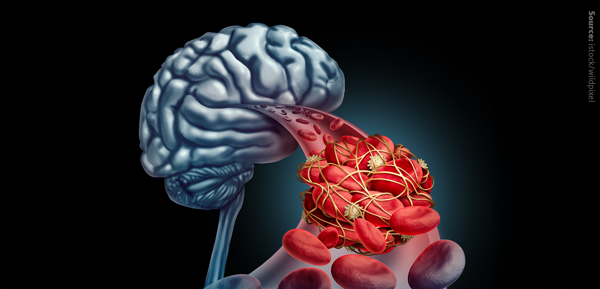
STROKE, one of the leading causes of mortality and disability worldwide, is characterized by the sudden onset of neurological deficits resulting from the interruption of blood supply to the brain. Ischemic stroke, which accounts for approximately 85% of all strokes, occurs when a clot or plaque obstructs a cerebral artery, depriving brain tissue of oxygen and nutrients. This leads to rapid cell death, neuronal injury and an inflammatory response.
Inflammation plays an important role in determining the extent of neuronal damage and recovery. At the centre of this inflammatory response are microglia – the brain’s immune cells. Microglia, while essential for managing brain injury with a controlled inflammatory response, can worsen brain damage when their activity becomes dysregulated.

The recent research by Wei Wei and colleagues sheds light on the involvement of lipid droplets and lipid metabolism in microglial function post-ischemic stroke, revealing its potential as a therapeutic target.
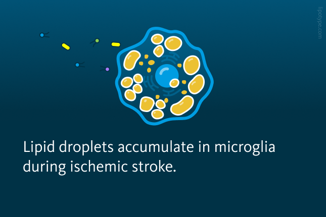
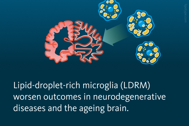
More and more evidence points towards lipid droplet accumulation in microglia during hypoxic conditions, for example in the case of ischemic stroke. Lipid droplets are specialized organelles that store lipids including cholesterol (Chol), cholesteryl esters (CE) and triglycerides (TAG). These lipid droplets, though vital for maintaining cellular energy and lipid balance, seem to drive microglia into a dysfunctional, pro-inflammatory state. This phenotype, referred to as lipid-droplet-rich microglia (LDRM) has been linked to worsened outcomes in ageing brains and neurodegenerative diseases. This study investigates the dynamic interactions between lipid metabolism, microglial phenotypes, and inflammation in ischemic stroke models to provide insights into the mechanisms driving stroke-induced neuroinflammation.

In ischemic stroke models, microglia exposed to hypoxia and inflammation showed a marked increase in lipid droplet biogenesis and changes in lipid metabolism. This was evident both in vitro and in vivo models with lipid droplet accumulation.
In vitro experiments using primary microglia cells exposed to hypoxic or inflammatory conditions revealed significant lipid droplet accumulation. Hypoxia-induced lipid droplet formation in microglia peaked after 24 hours of reoxygenation, and this accumulation was further enhanced by inflammatory stimuli, such as lipopolysaccharide. Interestingly, inhibiting Acyl-CoA acyltransferase (ACAT), an enzyme involved in lipid metabolism, significantly reduced lipid droplet accumulation, suggesting that lipid metabolism plays a crucial role in regulating microglial activation under ischemic conditions.
 accumulation in hypoxia. A Bar graph showing the increased amount of LDRM in hypoxia conditions in cultured microglia. B The boxplot shows the increase in LDRM for different durations of hypoxia conditions. The longer the microglia are exposed, the more LDRM are forming, peaking at 24 h.")
Lipid droplet-rich microglia (LDRM) accumulation in hypoxia. A Bar graph showing the increased amount of LDRM in hypoxia conditions in cultured microglia. B The boxplot shows the increase in LDRM for different durations of hypoxia conditions. The longer the microglia are exposed, the more LDRM are forming, peaking at 24 h.
Wei Wei et al. Advanced Science, 11.41: 2306863 (2024), 10.1002/advs.202306863
In vivo, using rodent models of middle cerebral artery occlusion (MCAO), researchers observed a temporal and spatial pattern of lipid droplet accumulation in ischemic brain regions, particularly peaking from 3 to 7 days post-stroke. Based on FACS measurements, MCAO-induced lipid droplet formation was apparent mainly in the lesioned hemisphere, particularly within cortical and subcortical brain regions. These LDRM exhibited elevated levels of inflammatory markers such as TNF-α, IL-1β, and IL-6, alongside downregulation of anti-inflammatory markers like IL-10. In co-culture systems, neurons exposed to LDRMs demonstrated increased vulnerability, emphasizing the detrimental effects of this microglial phenotype.
One of the most striking discoveries was the spatial and temporal heterogeneity in lipid composition across different brain regions poststroke. Using the MCAO mouse model, researchers analyzed lipid profiles in the ischemic core and surrounding areas. Shotgun mass spectrometry lipidomics was performed on tissue samples from different brain regions at various time points post-stroke. For this purpose, six regions within the ischemic brain were defined: the ipsilateral cortex within the core of the lesion (L Core), the ipsilateral cortex outside of the lesion (L Cortex), the white matter at the edge of the ischemic lesion (L Edge), and the corresponding regions on the contralateral side (NL Core, NL Corte, and NL Edge). Initial data exploration of lipid metabolism by principal component analysis (PCA) of sham, days 3 and 7 post-ischemia in each of the abovementioned brain regions revealed that in addition to the regional separation, the 3 d L Core and the 7 d L Core appeared to have distinctive lipid profiles.
.")
Brain regions of interest in MCAO mouse model for lipidomics. Definition of regions of interest on MCAO-induced mouse brain tissue; (here the definitions of Rs and Ls).
Wei Wei et al. Advanced Science, 11.41: 2306863 (2024), 10.1002/advs.202306863
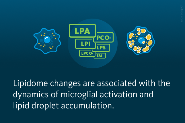
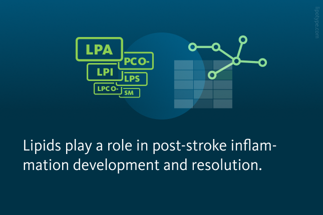
The lipidomic analysis of brain regions with ischemic stroke revealed distinct lipidomic changes, with the core region showing a significant increase in cholesteryl esters, TAGs, and diacylglycerols , particularly at seven days post-ischemia. Furthermore, lipidomic analysis highlighted dynamic changes in phosphatidylcholine (PC), phosphatidylethanolamine (PE), and their ether-linked variants (PC O-, PE O-) during the early post-stroke stages. These findings suggest that localized lipid alterations may directly influence microglial activation states and inflammatory responses.
 of lipid composition patterns in different regions of brains (sham, post-ischemia days 3 and 7). B Heatmap of core regions representing color-coded Z scores of lipid-class distributions of the 24 most abundant lipid classes using the lipid species concentrations (pmol) with log2 normalization.")
Lipid-class composition of distinct mouse brain regions after stroke. A Principal component analysis (PCA) of lipid composition patterns in different regions of brains (sham, post-ischemia days 3 and 7). B Heatmap of core regions representing color-coded Z scores of lipid-class distributions of the 24 most abundant lipid classes using the lipid species concentrations (pmol) with log2 normalization.
Wei Wei et al. Advanced Science, 11.41: 2306863 (2024), 10.1002/advs.202306863
These lipid changes in the brain with ischemic stroke were closely associated with the dynamics of microglial activation and lipid droplet accumulation. In particular, the lipid composition in the core region correlated with the presence of pro-inflammatory microglia, while the edge region exhibited a more anti-inflammatory phenotype with reduced lipid accumulation. This suggests that lipid metabolism and microglial phenotype are tightly linked and that changes in lipid profiles could influence the inflammatory response and tissue damage after ischemic injury.
This study provides insights into the role of lipid metabolism and lipid droplet accumulation in microglia during ischemic stroke. Ischemia induces dynamic alterations in brain lipid profiles, which in turn modulate the microglial phenotype and inflammatory response. While this study focuses on ischemic stroke, the implications of lipid metabolism in neuroinflammation extend to other conditions, including neurodegenerative diseases including Alzheimer’s disease and Parkinson’s disease. The ability of microglia to transition between pro- and anti-inflammatory states is heavily influenced by their lipid environment. By understanding and manipulating these lipid dynamics, it may be possible to develop interventions that restore microglial homeostasis across a range of neurological disorders.
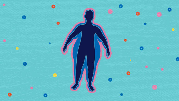
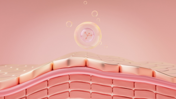
Lipotype Lipidomics allows for precise and comprehensive analysis of lipid droplets. By profiling the lipid content of these structures, it is possible to detect changes in lipid metabolism associated with conditions such as obesity, diabetes, cardiovascular diseases, and neurodegenerative disorders.
Not sure how this works for your case?
Let’s talk it through!

Lipotype products are provided for Research Use Only. They are not intended for clinical diagnostic purposes and must not be used to inform medical treatment decisions. The content of this article is for scientific and educational purposes only and should not be considered medical advice.