
INFLAMMATORY bowel disease (IBD) is a condition that causes inflammation in the intestines, either in scattered inflammation areas throughout the digestive tract (Crohn’s disease) or continuously in the colon (ulcerative colitis).
Cytokines play a crucial role in the development of IBD. They regulate the immune response by controlling inflammation and T-cell differentiation in the gut. An imbalance between pro-inflammatory cytokines (TNF-α, IL-1, IL-6, IL-17) and anti-inflammatory ones (IL-10, IL-4) leads to chronic inflammation and tissue damage.
 is a condition that causes inflammation in the intestines, either in scattered inflammation areas throughout the digestive tract (Crohn’s disease) or continuously in the colon (ulcerative colitis).")
TNF-α is a major pro-inflammatory cytokine in IBD, driving inflammation by triggering other cytokines, impacting cell growth, apoptosis, and immune processes, which has a negative effect on the colonic environment. High TNF-α levels in the inflamed intestinal tissues correlate with the disease severity.
While anti-TNF-α antibody drugs are available, since IBD mainly affects intestinal tissues, the non-targeted delivery mechanism of these therapies in application to IBD can lead to systemic absorption, reducing effectiveness and causing systemic side effects. Monoclonal antibodies must also recognize complex protein structures, limiting their ability to precisely target molecules. In contrast, siRNA therapies offer a promising alternative, as they can selectively target any gene by matching the correct nucleotide sequence to the target mRNA. Oral drug delivery is in general preferred as it allows direct treatment at the affected site in the intestines.

The oral delivery of biologic drugs, especially RNA-based therapies, is challenging due to their vulnerability to the gastrointestinal environment and difficulty crossing the intestinal barrier. To overcome these issues, formulation-based delivery technologies are needed to enhance drug stability and absorption. To address this, Geonhee Han and colleagues developed an exosome-based platform for siRNA delivery, designed to treat inflammatory bowel disease by targeting inflamed colonic sites via oral administration and modulating the immune response. Exosomes, naturally involved in intercellular communication and macromolecule transport, offer a promising targeted drug delivery system.
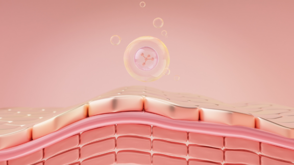
Milk-derived exosomes are a unique class of extracellular vesicles that protect packaged biomolecules as they pass through the stomach and gastrointestinal tract. Unlike exosomes from mammalian cells, which degrade under harsh conditions, milk-derived exosomes remain stable and can be absorbed by intestinal and colonic cells. This makes them an effective vehicle for oral drug delivery in the treatment of diseases like inflammatory bowel disease. Milk-derived exosomes are highly resistant to digestive enzymes and pH variations, allowing them to deliver molecules to various organs, including the liver, brain, placenta, and gut.

In this study, the authors used electroporation to load TNF-α siRNA into milk-derived exosomes and tested its effectiveness in treating colitis in mice. They explored whether delivering TNF-α siRNA via milk-derived exosomes through oral gavage could help reduce colitis symptoms. The experiment involved inducing colitis with dextran sulfate sodium (DSS) and administering TNF-α siRNA via milk-derived exosomes through intraperitoneal injections. After an acclimatization period, mice received four intraperitoneal injections of milk-derived exosomes with TNF-α siRNA at three-day intervals, starting one day before being given DSS.
 and administering TNF-α siRNA via milk-derived exosomes through intraperitoneal injections. After an acclimatization period, mice received four intraperitoneal injections of milk-derived exosomes with TNF-α siRNA at three-day intervals, starting one day before being given DSS.")
Further experiments confirmed that exosomes keep their structure after electroporation, ensuring full siRNA encapsulation milk-derived exosomes with TNF-α siRNA help reduce inflammation by lowering TNF-α levels without causing cell toxicity. Effective siRNA delivery minimizes colitis symptoms and histological manifestation by suppressing TNF-α expression. When given orally, TNF-α siRNA in milk-derived exosomes successfully reaches the intestines. Oral administration of milk-derived exosomes with TNF-α siRNA helps restore the balance of inflammatory cytokines disrupted by colitis. In case of intraperitoneal administration of milk-derived exosomes with TNF-α siRNA, a mild loss of the surface epithelium was observed compared to drastic changes in surface epithelium without treatment.

To assess siRNA delivery capacity, researchers evaluated the physicochemical properties of exosomes by analyzing the lipid composition of exosomes. They compared milk-derived exosomes and HEK293T cells-derived exosomes. Cell-derived exosomes primarily contained phosphatidylcholine (PC), ether-linked phosphatidylethanolamine (PE O-), phosphatidylserine (PS), cholesterol esters (CE), and ether-linked phosphatidylcholine (PC O-) lipids. In contrast, milk-derived exosomes were rich in triglyceride (TAG), phosphatidylethanolamine (PE), diacylglycerol (DAG), and PC lipid classes, with TAG comprising over half of their lipid content.
Notably, milk-derived exosomes had a significantly lower PC/PE lipid ratio than cell-derived exosomes. This unique lipid composition contributes to their exceptional stability in the gastrointestinal tract. The high PE content thickens the membrane, enhancing stability compared to cell-derived exosomes, as indicated by the PC/PE ratio, which is a key factor in membrane dynamics.

Lipid composition of milk-derived exosomes and cell-derived exosomes. A Major lipid classes measured in exosomes. B Lipid classes distribution in milk-derived and cell-derived exosomes.
Han et al., Bioactive Materials, 34, 138-149 (2024), 10.1016/j.bioactmat.2023.12.010
From a lipidomics perspective, milk-derived exosomes demonstrate exceptional stability due to their unique lipid composition, distinguishing them from cell-derived exosomes. After oral administration, milk-derived exosomes effectively pass through the gastrointestinal tract and reach colitis lesions, where they prompt the degradation of TNF-α mRNA.
This study confirms that suppressing TNF-α expression improves IBD symptoms, allowing the researchers to claim that milk-derived exosomes are a robust tool for oral gene therapy in colitis. The distinct membrane composition of milk-derived exosomes ensures their stability through the gastrointestinal tract, allowing efficient delivery to inflamed lesions, where they degrade TNF-α mRNA. This reduction in TNF-α expression lowers pro-inflammatory cytokine and reactive oxygen species levels, supporting the treatment of intestinal colitis.


Lipotype Lipidomics technology helps to study the structural and functional roles of lipids in biological systems, supporting the development of targeted drug delivery, disease biomarkers, and novel therapeutic strategies across various medical fields.
Not sure how this works for your case?
Let’s talk it through!

Lipotype products are provided for Research Use Only. They are not intended for clinical diagnostic purposes and must not be used to inform medical treatment decisions. The content of this article is for scientific and educational purposes only and should not be considered medical advice.