CELLS continuously internalize molecules from their environment by folding their outer membrane inward, forming small transport vesicles that carry cargo into the cell, a process known as endocytosis. These vesicles first fuse with early endosomes, where cargo is sorted: some molecules are recycled back to the cell surface, while others continue deeper into the cell. Material is also delivered to endosomes from the trans-Golgi network – the cell’s sorting station for newly made proteins and lipids – where molecular signals determine whether cargo continues toward lysosomes for degradation or is returned to the Golgi for further processing.
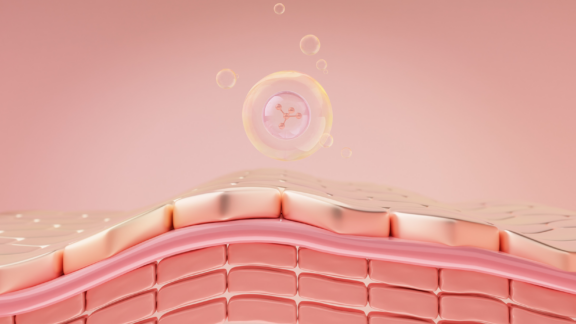
Cargo destined for degradation eventually reaches late endosomes, which mature into lysosomes. These are the cell’s primary recycling compartments, responsible for breaking down waste materials and releasing their molecular building blocks for reuse. Together, these late endosomes and lysosomes form the cell’s core lipid processing hub.

NPC1 and NPC2 are proteins that work together to transport cholesterol and other lipids through the cell’s recycling system. NPC1 is a large membrane-spanning protein embedded in the wall of the late endosome and lysosome compartments, while NPC2, a smaller soluble protein, works in tandem by binding cholesterol inside the lysosome and handing it over to NPC1 for export out of the compartment. When either protein is absent or dysfunctional, this lipid export system fails, and cholesterol, along with glycolipids, accumulates to toxic levels within late endosomes and lysosomes, unable to reach the rest of the cell where they are needed.
The consequences of this lipid trafficking failure extend far beyond the lysosome itself. Cholesterol is not just a structural building block of cell membranes; it is also an essential precursor for steroid hormones, including neurosteroids, which are critical for brain function. When cholesterol is trapped inside lysosomes, it becomes unavailable for these vital roles elsewhere in the cell. At the same time, the buildup of glycosphingolipids in the nervous system triggers abnormal structural changes in neurons, disrupting their architecture and connectivity. Together, these molecular disruptions create a cascade of cellular dysfunction that drives the progressive neurological decline seen in Niemann-Pick disease type C (NPC) patients.
. (<strong>A</strong>) In healthy cells, lipoprotein particles deliver cholesterol to the cell via surface receptors, after which it enters the late endosomal and lysosomal system. The NPC1 and NPC2 proteins then ensure its safe passage to the Golgi and endoplasmic reticulum for further use. (<strong>B</strong>) Loss of NPC1 or NPC2 function breaks this transport route. Unesterified (free) cholesterol accumulates inside late endosomes and lysosomes, starving other cellular compartments of this essential molecule.")
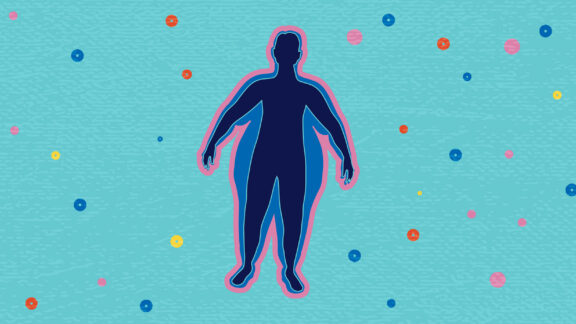
Niemann-Pick disease type C is a rare, inherited lysosomal storage disease caused by mutations in the NPC1 or NPC2 genes (Niemann-Pick disease by Mayo Clinic, Niemann-Pick disease Type C by EffRx Pharmaceuticals). These genetic defects disrupt the intracellular transport of cholesterol and other lipids, leading to their toxic accumulation in the liver, spleen, lungs, and the central nervous system. NPC affects individuals across all age groups, with symptoms ranging from neonatal liver disease to adult-onset neurodegeneration.
The neurological progression of NPC is particularly debilitating. As lipids accumulate in neurons over time, patients develop progressive loss of motor control, impaired eye movements, difficulties with speech and swallowing, and cognitive decline. Currently, there is no cure for NPC. Disease management remains largely supportive, though emerging therapeutic strategies – including substrate reduction and lipid-targeting approaches – are reshaping the treatment landscape.

Since NPC1 dysfunction originates within a specific intracellular compartment – the late endosome and lysosome – understanding the disease requires tools that can zoom in at the same level of precision. Omics approaches such as proteomics and lipidomics offer powerful ways to map the full molecular landscape of a diseased cell, revealing disrupted metabolic and signalling pathways that would otherwise go undetected. When applied to whole cells or tissues, these approaches face a fundamental challenge: the complexity of thousands of proteins and lipids distributed across all cellular compartments at once can easily mask the subtle but critical changes occurring in one specific location. The solution lies in subcellular fractionation – isolating individual compartments before analysis. Yet not all organelles are equally easy to isolate. Highly structured compartments such as mitochondria, synaptic vesicles, and centrosomes can be purified with relatively high yield and consistency thanks to their distinct physical properties.
The dynamic nature and physicochemical overlap of late endosomes and lysosomes with other membrane structures make conventional isolation strategies unreliable. This raises the risk that contaminants from neighboring compartments could affect omics results or incorrectly assign molecular signals to the wrong cellular location. To overcome this, Tharkeshwar and colleagues employed a magnetic isolation strategy using superparamagnetic iron oxide nanoparticles (SPIONs). This approach uses the unique magnetic properties of nanoparticles to selectively capture and purify the late endosomes and lysosomes compartments with high specificity. While SPIONs have been widely explored in biomedical applications such as drug delivery, cancer hyperthermia treatment, and MRI contrast enhancement, their use as a precision tool for organelle isolation represents a new application that is particularly suited to resolving the proteomics and lipidomics complexity of NPC1.
, arranged around a central illustration of a SPION particle. The applications are: bioimaging and biosensors, drug delivery, enzyme immobilization, hyperthermia, organelle and cell compartment separation, toxin removal, tissue engineering, and cell labelling and sorting.")
To isolate distinct subcellular compartments, the researchers engineered SPIONs with tailor-made surface coatings, directing each nanoparticle type to a specific cellular destination. Aminolipid-coated SPIONs associated with and remained at the plasma membrane, while DMSA-coated SPIONs were internalized by the cell and trafficked to late endosomes and lysosomes. Both coating strategies produced stable superparamagnetic nanoparticles small enough to interact with cellular structures while retaining the magnetic properties essential for downstream isolation. This differential targeting allowed the researchers to physically separate the plasma membrane and late endosome and lysosome compartments from the same cells using a magnetic workflow.
The successful separation of compartments was confirmed through omics profiling, which revealed distinct, compartment-specific molecular signatures for each isolated fraction. The authors then applied this methodology to both wild-type and NPC1-deficient HeLa cells (a well-established cellular model of NPC), allowing for a compartment-resolved lipidome and proteome comparison. This way, scientists evaluated how NPC1 loss reshapes the molecular landscape of the late endosomes and lysosomes.
 used in this study. Both particles share the same iron oxide core. The DMSA-coated SPION (left, shown in red, 10nm) has a compact appearance, with short wavy projections radiating closely around the core. The aminolipid-coated SPION (right, shown in yellow, 16nm) appears larger overall, with the core first wrapped in a soft layer, then surrounded by long, loose, wavy chains extending outward in all directions.")
With compartment purity established by proteomics analysis, the researchers applied shotgun lipidomics to map lipid changes across whole cells, plasma membranes, and late endosomes and lysosomes in wild-type versus NPC1-deficient cells. They detected, identified, and quantified 551 individual lipid species across 17 lipid classes. At the whole-cell level, NPC1-deficient cells showed an increase in total cholesterol, which was consistent with the hallmark cholesterol transport blockade in NPC. Interestingly, the plasma membrane lipidome remained largely unchanged between wild-type and NPC1-deficient cells, suggesting that lipid disruption is highly compartment-specific. The most pronounced changes were concentrated in the late endosomes and lysosomes fraction: glycerophospholipids were significantly reduced, while cholesterol and storage lipids accumulated strongly, together accounting for over 50 mol% of the lysosomal lipidome in NPC1-deficient cells compared to much lower levels in wild-type.
, plasma membrane (PM), and late endosomes and lysosomes (LYS) in control and NPC1-deficient cells. The lipid classes shown are glycerophospholipids (GPL) (PC, PE, PI, PS, PG, PC O-, PE O-, PA, DAG, sphingolipids (SL), storage lipids including triacylglycerols and cholesterol esters (TAG+CE), cholesterol (Chol), and others. While the lipid composition of TCL and PM fractions remains broadly similar between control and NPC1-deficient cells, the late endosomes and lysosomes fraction shows pronounced differences. In NPC1-deficient lysosomes, glycerophospholipids decrease from 59.6% to 41.2%, while cholesterol rises from 28.0% to 37.9% and storage lipids increase markedly from 4.6% to 15.3%. Tharkeshwar et al., Sci Rep. 2017;7:41408. 10.1038/srep41408")
Lipid composition changes across cellular compartments in NPC1-deficient cells. The mole percentage of major lipid classes is compared across three cellular fractions, namely total cell lysate (TCL), plasma membrane (PM), and late endosomes and lysosomes (LYS) in control and NPC1-deficient cells. The lipid classes shown are glycerophospholipids (GPL) (PC, PE, PI, PS, PG, PC O-, PE O-, PA, DAG, sphingolipids (SL), storage lipids including triacylglycerols and cholesterol esters (TAG+CE), cholesterol (Chol), and others. While the lipid composition of TCL and PM fractions remains broadly similar between control and NPC1-deficient cells, the late endosomes and lysosomes fraction shows pronounced differences. In NPC1-deficient lysosomes, glycerophospholipids decrease from 59.6% to 41.2%, while cholesterol rises from 28.0% to 37.9% and storage lipids increase markedly from 4.6% to 15.3%.
Tharkeshwar et al., Sci Rep. 2017;7:41408. 10.1038/srep41408
Nearly all major glycerophospholipid classes, including phosphatidylcholine (PC), phosphatidylethanolamine (PE), phosphatidylserine (PS), and phosphatidylinositol (PI), as well as their ether-linked variants, showed more than three-fold increases in the late endosomes and lysosomes of NPC1-deficient cells. Sphingolipids, including ceramides (Cer), sphingomyelins (SM), and hexosylceramides (HexCer), were also significantly elevated. The simultaneous accumulation of both ceramide and cholesterol in the same compartment is unexpected, as ceramide is known to displace cholesterol from membranes. Their co-accumulation in the NPC1-deficient lysosome suggests a profound disruption of lipid homeostasis rather than a simple storage defect. Particularly striking was a greater than ten-fold increase in lysophosphatidylcholines (LPC) in the late endosomes and lysosomes fraction. This change is not observed in whole-cell lysates or plasma membranes, which underlines that these lipid alterations are compartment-specific.
Beyond phospholipids, glycosphingolipid levels were also significantly elevated in both whole-cell lysates and late endosomes and lysosomes fractions of NPC1-deficient cells, consistent with the higher ceramide levels observed, since ceramide serves as the key precursor for glycosphingolipid synthesis. Interestingly, while total glycosphingolipid increases were pronounced at the whole-cell level, the rise within the lysosomal fraction itself was more moderate, suggesting that glycosphingolipid accumulation is not confined to the lysosome but reflects a broader disruption of sphingolipid metabolism across the cell.
, sphingolipids (ceramide, hexosylceramide and sphingomyelin), storage lipids (triacylglycerols and cholesterol esters), cholesterol, and lyso-lipid species. The dashed line at 1 represents the control level. Individual lipid species Lac, Gb3, Gb4, pGb, GA2, GM2 and GM3, are elevated in the late endosome and lysosome fraction of both NPC1-knockout lines compared to controls, with Gb3 showing the most pronounced increase. Total GSL levels are markedly higher in NPC1-deficient late endosomes and lysosomes. Tharkeshwar et al., Sci Rep. 2017;7:41408. 10.1038/srep41408")
Lipid accumulation is specific to late endosomes and lysosomes in NPC1-deficient cells. Relative fold changes in lipid class abundance are shown for the lysosomal fraction of two independent NPC1-knockout cell lines compared to control cells. Major lipid classes are elevated, including glycerophospholipids (PC, PE, PI, PS, PG and their ether-linked forms, PA and DAG), sphingolipids (ceramide, hexosylceramide and sphingomyelin), storage lipids (triacylglycerols and cholesterol esters), cholesterol, and lyso-lipid species. The dashed line at 1 represents the control level. Individual lipid species Lac, Gb3, Gb4, pGb, GA2, GM2 and GM3, are elevated in the late endosome and lysosome fraction of both NPC1-knockout lines compared to controls, with Gb3 showing the most pronounced increase. Total GSL levels are markedly higher in NPC1-deficient late endosomes and lysosomes.
Tharkeshwar et al., Sci Rep. 2017;7:41408. 10.1038/srep41408
Proteomic analysis of isolated late endosomes and lysosomes fraction revealed that NPC1 deficiency triggers broad molecular remodelling specifically within the late endosomes and lysosomes compartment, while the plasma membrane proteome remains largely unaffected, mirroring the compartment-specific lipid changes observed in lipidomics. Among the 53 differentially expressed lysosomal proteins identified, key enzymes involved in lipid metabolism were downregulated, while proteins linked to autophagy and lysosomal biogenesis were strongly upregulated. This suggests that cells attempt to compensate for lysosomal dysfunction by ramping up their degradation machinery. Despite this compensatory response, lysosomal enzymatic activity was paradoxically reduced in NPC1-deficient cells, pointing to a fundamental impairment in lysosomal function that goes beyond simple lipid storage.
This research demonstrates how the combination of SPION-based organelle isolation with high-resolution lipidomics can reveal the molecular fingerprint of a rare lysosomal storage disorder with subcellular precision. By isolating and analyzing the compartment where NPC1 dysfunction originates, researchers identified a highly compartment-specific pattern of lipid dysregulation that a whole-cell approach alone would have missed. This approach provides new molecular insights into the pathology of Niemann-Pick disease type C1 and highlights the late endosome and lysosome compartment as a central hub of disease-driven molecular remodelling.


Lipotype Lipidomics technology is a powerful tool to study lysosomal storage disorders. Profiling lipid composition at the subcellular level pinpoints which lipid classes are disrupted, in which compartments, and to what extent. Beyond lysosomal storage diseases, this approach holds broad potential across rare diseases involving dysregulated lipid metabolism, from biomarker discovery to the identification of novel therapeutic targets.
Need clarity on the process?
Ask us anything!

Lipotype products are provided for Research Use Only. They are not intended for clinical diagnostic purposes and must not be used to inform medical treatment decisions. The content of this article is for scientific and educational purposes only and should not be considered medical advice.